Every time a cell divides, repairs damaged DNA, or defends itself against invading viruses, it relies on a class of enzymes most people have never heard of: nucleases.
These molecular scissors cut nucleic acids, but not all nucleases work the same way. Two major types and endonucleases differ fundamentally in where and how they cut, and that difference shapes everything from DNA replication to modern gene-editing technology.
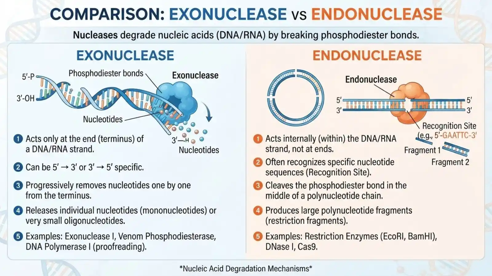
Exonucleases trim nucleotides one at a time from the ends of a DNA or RNA strand, working like a zipper being slowly undone.
Endonucleases, by contrast, cut internally, slicing the strand at specific points along its length, much like scissors cutting through the middle of a ribbon.
What Are Nucleases?
Nucleases are a broad class of enzymes that break down nucleic acidsDNA and RNAby targeting the phosphodiester bonds that link individual nucleotides together into a chain.
Without these bonds, a nucleic acid strand would simply fall apart into its component pieces. Nucleases exploit this by selectively hydrolyzing specific bonds, allowing cells to cut, trim, degrade, or repair genetic material as needed.
These enzymes are essential across nearly every biological process involving DNA or RNA. They help cells copy their genome accurately, remove damaged or incorrect sequences, recycle old genetic material, and defend against foreign DNA from viruses or invading organisms. Without nucleases, processes like DNA replication, repair, and immune defense simply couldn’t function.
Nucleases are typically classified along two main axes. The first is substrate specificity: DNases act on DNA, while RNases act on RNA, though some nucleases can process both. The second, more functionally important axis is cleavage location, which divides nucleases into two major groups: exonucleases and endonucleases.
This exo/endo distinction isn’t just a technical detail it reflects fundamentally different mechanisms, structural designs, and biological roles. An enzyme built to recognize a free strand end looks and behaves very differently from one designed to locate and cut a specific internal sequence.
The discovery of these enzyme classes, dating back to foundational molecular biology research in the mid-20th century, paved the way for tools like restriction enzymes and CRISPR that now define modern biotechnology.
Exonucleases: Definition and Mechanism
Exonucleases are enzymes that cleave nucleotides one at a time from the terminal ends of a DNA or RNA strand.
Unlike enzymes that attack the middle of a sequence, exonucleases require a free end to initiate cutting, making them incapable of acting on circular DNA molecules that lack accessible termini unless that circle is first nicked or broken.
A defining feature of exonucleases is directionality. Some enzymes work in the 5′ to 3′ direction, removing nucleotides starting from the strand’s 5′ end and moving toward the 3′ end. Others work 3′ to 5′, doing the reverse. This directionality isn’t arbitrary; it’s dictated by the enzyme’s active site architecture, which is shaped to recognize and bind a specific terminal chemistry.
Structurally, exonucleases possess active sites that clamp onto the very end of a nucleic acid strand, positioning the terminal nucleotide for hydrolysis before releasing it and shifting to the newly exposed end. This creates a stepwise, sequential cutting pattern rather than a single internal break.
Exonucleases also vary in processivity. Highly processive exonucleases remain bound to the same strand, chewing through many nucleotides in a continuous run before dissociating. Distributive exonucleases, on the other hand, release the strand after removing just one or a few nucleotides, requiring frequent rebinding to continue.
This end-specific, directional cutting mechanism makes exonucleases particularly suited for tasks like proofreading during DNA replication, where errors near a growing strand’s end need to be trimmed away, and for degrading unwanted or damaged nucleic acid fragments.
Endonucleases: Definition and Mechanism
Endonucleases are enzymes that cleave phosphodiester bonds within the interior of a DNA or RNA strand, rather than at its ends.
Unlike exonucleases, they don’t need a free terminus to act, they can recognize and cut internal sites even on circular DNA molecules like plasmids, which have no accessible ends at all.
The defining feature of endonucleases is target recognition. Some, like many restriction enzymes, are highly sequence-specific, scanning DNA until they locate a particular recognition sequenceoften a short palindromic motifbefore making a precise cut. Others are non-specific, cleaving essentially anywhere along the strand, which is useful for broadly degrading nucleic acids rather than targeting a defined site.
Structurally, endonucleases have active sites built to insert into or wrap around the nucleic acid strand at an internal position, rather than clamping onto a terminal end. Many sequence-specific endonucleases work as dimers, with two identical subunits each cutting one strand of double-stranded DNA at matching positions.
This internal cutting produces a defining structural outcome: the type of end generated at the cleavage site. Blunt-end cutters slice both strands at the same position, leaving flush ends. Sticky-end cutters make staggered cuts, leaving short single-stranded overhangs that can later base-pair with complementary overhangs, a property heavily exploited in molecular cloning.
Because they can act anywhere along a strand rather than only at termini, endonucleases are essential for tasks like introducing double-strand breaks for repair pathways, initiating excision repair, and enabling programmable gene editing tools like CRISPR-Cas9.
Side-by-Side Comparison
While both enzyme classes cleave phosphodiester bonds, the way they do soand the biological logic behind it differs substantially. The table below summarizes the core distinctions.
| Feature | Exonucleases | Endonucleases |
| Cleavage site | Terminal ends only | Internal positions |
| Requires free end? | Yes | No |
| Acts on circular DNA? | No (unless nicked first) | Yes |
| Directionality | 5’→3′ or 3’→5′ | No inherent directionality |
| Specificity | Often less sequence-specific | Often highly sequence-specific |
| Cutting pattern | Sequential, one nucleotide at a time | Single or multiple internal cuts |
| Products | Mononucleotides | Larger DNA/RNA fragments |
| Common examples | Exonuclease I, Exonuclease III, Lambda exonuclease | EcoRI, DNase I, Cas9 |
Beyond mechanism, substrate preference also varies. Some exonucleases and endonucleases act exclusively on single-stranded nucleic acids, others only on double-stranded molecules, and some can process both depending on cellular context. This flexibility allows cells to deploy the right enzyme for the right situation whether that’s trimming a single mismatched base or excising an entire damaged segment.
The products generated also differ meaningfully. Exonucleases typically release individual nucleotides as their end product, effectively recycling raw material for the cell. Endonucleases, in contrast, generate fragments of varying length, which often serve as substrates for downstream enzymes such as ligases that reseal breaks or polymerases that fill gaps.
Key Examples of Exonucleases
Several well-characterized exonucleases illustrate the diversity of this enzyme class across different organisms and biological contexts.
Exonuclease I (E. coli) degrades single-stranded DNA in the 3′ to 5′ direction. It’s widely used in molecular biology to remove leftover single-stranded primers after PCR, particularly in Sanger sequencing cleanup protocols.
Exonuclease III (E. coli) removes nucleotides from the 3′ end of double-stranded DNA, and is commonly used in lab techniques to generate unidirectional deletions or create single-stranded regions for downstream cloning applications.
DNA Polymerase I’s proofreading activity is a built-in 3′ to 5′ exonuclease function. As the polymerase synthesizes new DNA, this activity immediately detects and excises mismatched nucleotides, dramatically increasing replication fidelity.
Exonuclease V, part of the RecBCD complex in bacteria, combines both exonuclease and helicase activity. It unwinds and degrades DNA at double-strand breaks, playing a central role in homologous recombination repair.
Lambda exonuclease, derived from bacteriophage lambda, degrades DNA in the 5′ to 3′ direction and is a staple tool in molecular cloning, particularly for generating single-stranded DNA templates.
Poly(A)-specific ribonuclease (PARN) illustrates that exonuclease activity isn’t limited to DNAit trims the poly(A) tail from mRNA molecules, a key step in regulating mRNA stability and turnover.
Key Examples of Endonucleases
Endonucleases span an equally diverse range of functions, from bacterial defense systems to programmable gene editing.
Restriction endonucleases (EcoRI, HindIII, BamHI) are perhaps the most famous examples. Originally evolved as part of bacterial immune systems to destroy invading viral DNA, these enzymes recognize specific short palindromic sequences and cut at precise positions. Their predictable cutting patterns made them foundational tools in molecular cloning, allowing scientists to cut and paste DNA fragments decades before CRISPR existed.
DNase I is a relatively non-specific endonuclease that cleaves DNA at essentially any accessible internal site. It’s widely used in labs to degrade DNA contamination in RNA preparations and in chromatin accessibility assays like DNase-seq.
CRISPR-Cas9 functions as an RNA-guided endonuclease. Guided by a short RNA sequence complementary to its target, Cas9 introduces a precise double-strand break at a specific genomic location, forming the basis of modern gene editing.
Ribonucleases like RNase A and RNase H also act as endonucleases on RNA substrates. RNase H specifically degrades the RNA strand within an RNA-DNA hybrid, a function essential in processes like Okazaki fragment processing during replication.
AP endonucleases play a critical role in base excision repair, recognizing basic sites/positions where a damaged base has already been removed and cutting the DNA backbone there to allow repair machinery to complete the fix.
Biological Roles and Pathways
Exonucleases and endonucleases aren’t just isolated biochemical curiosities; they’re embedded in nearly every core pathway that maintains genomic integrity and cellular function.
DNA replication relies heavily on both enzyme types. Proofreading exonucleases built into DNA polymerases correct misincorporated bases in real time, while endonucleases help process Okazaki fragments on the lagging strand, removing RNA primers so they can be replaced with DNA and sealed by ligase.
DNA repair pathways depend on precise coordination between the two. In nucleotide excision repair, endonucleases make incisions on either side of a DNA lesion, after which exonuclease-like activity removes the damaged segment for resynthesis. Mismatch repair follows a similar logic, using endonucleases to nick the strand and exonucleases to excise the surrounding error-containing region.
RNA processing and degradation also depend on both enzyme classes. Exonucleases trim mRNA poly(A) tails and degrade transcripts during turnover, while endonucleases make internal cuts that initiate decay pathways or process precursor RNAs into mature rRNA and tRNA molecules.
Host defense mechanisms in bacteria showcase endonucleases prominently, particularly restriction-modification systems that destroy foreign viral DNA, and CRISPR-Cas systems that provide adaptive immunity against future infections.
Apoptosis, or programmed cell death, relies on endonucleases to fragment genomic DNA into characteristic pieces, a hallmark used by researchers to confirm this form of cell death is occurring.
Biotechnology and Research Applications
Beyond their natural biological roles, exonucleases and endonucleases have become indispensable tools across modern biotechnology, powering techniques that define contemporary molecular biology.
Molecular cloning was revolutionized by restriction endonucleases, which allow scientists to cut DNA at precise sequences and insert foreign genes into plasmid vectors. This cut-and-paste capability, paired with DNA ligase, formed the foundation of recombinant DNA technology decades before newer editing tools emerged.
Sanger sequencing cleanup commonly uses a combination of Exonuclease I and Shrimp Alkaline Phosphatase (marketed as ExoSAP-IT) to remove leftover primers and unincorporated nucleotides after PCR, ensuring cleaner sequencing reads.
Next-generation sequencing library prep relies on both enzyme types at various stages, including endonucleases for DNA fragmentation and exonucleases for trimming adapter sequences and processing fragment ends.
Gene editing technologies, most notably CRISPR-Cas9, TALENs, and zinc finger nucleases, all function as programmable endonucleases capable of introducing targeted double-strand breaks at specific genomic locations, enabling precise gene knockouts, insertions, or corrections.
PCR-based diagnostics frequently exploit the 5′ to 3′ exonuclease activity of Taq polymerase in TaqMan probe-based assays, where probe cleavage during amplification generates a fluorescent signal used to detect and quantify specific DNA sequences in real time.
Synthetic biology and DNA assembly techniques, such as Gibson assembly, depend on exonuclease “chew-back” activity to create complementary single-stranded overhangs that allow multiple DNA fragments to be seamlessly joined together in a single reaction.
Clinical and Disease Relevance
Beyond their roles in basic biology and biotechnology, nucleases also carry direct significance for human health, with mutations or dysregulation in these enzymes linked to a range of diseases.
TREX1, a 3′ to 5′ exonuclease responsible for clearing excess DNA fragments from the cytoplasm, illustrates this connection clearly. Mutations in the TREX1 gene are associated with Aicardi-Goutières syndrome, a rare autoimmune condition where accumulated DNA fragments trigger an inappropriate immune response, mimicking a chronic viral infection. TREX1 mutations have also been linked to lupus-like autoimmune symptoms in some patients.
Cancer biology frequently intersects with nuclease function, particularly through DNA repair pathways. Defects in genes like BRCA1 and BRCA2, which support homologous recombination repair alongside nucleases like those in the RecBCD-like MRN complex, impair the cell’s ability to accurately repair double-strand breaks. This repair deficiency contributes to genomic instability, a hallmark of many cancers, and has also been exploited therapeutically through treatments like PARP inhibitors.
Nucleases as therapeutic tools represent a rapidly growing area, particularly in gene therapy. Engineered nucleases, including CRISPR-Cas9, zinc finger nucleases, and TALENs, are being developed and clinically tested to correct disease-causing mutations directly at the genomic level, treat genetic blood disorders, and even engineer immune cells to fight cancer.
Common Misconceptions
Despite being foundational concepts in molecular biology, exonucleases and endonucleases are often oversimplified or misunderstood. Clearing up a few common misconceptions helps build a more accurate picture of how these enzymes actually work.
“All nucleases are non-specific.” While some nucleases, like DNase I, cut relatively indiscriminately, many others are highly sequence-specific. Restriction endonucleases, for instance, recognize precise palindromic sequences and will not cut DNA lacking that exact motif. CRISPR-Cas9 takes specificity even further, using RNA guide sequences to target virtually any user-defined genomic location.
“Exonucleases only degrade DNA.” This is a common oversimplification. While many well-known exonucleases act on DNA, plenty of others target RNA. Poly(A)-specific ribonuclease, for example, trims mRNA tails, and various exoribonucleases play essential roles in RNA turnover and quality control pathways.
“Restriction enzymes and endonucleases are the same thing.” Restriction endonucleases are simply one subtype within the broader endonuclease category. Other endonucleases, like DNase I, AP endonucleases, and Cas9, have no relationship to bacterial restriction-modification systems yet still qualify as endonucleases based purely on their internal cleavage mechanism.
“Exonucleases and endonucleases always work independently.” As covered in Section 9, many critical pathways like mismatch repair and homologous recombination require both enzyme types working in coordinated sequence, not as isolated, competing mechanisms.
How to Identify/Test Nuclease Activity
Confirming and characterizing nuclease activity in the lab requires specific experimental approaches, ranging from classic gel-based methods to modern fluorescence-based assays.
Gel electrophoresis-based cleavage assays remain one of the most straightforward and widely used techniques. A DNA or RNA substrate is incubated with the enzyme of interest, then run on an agarose or polyacrylamide gel. Exonuclease activity typically produces a gradual size shift or complete degradation into small fragments, while endonuclease activity often generates specific, discrete bands corresponding to predictable cut sites particularly useful when mapping restriction enzyme recognition sequences.
Fluorescence-based reporter assays offer a faster, more sensitive alternative. These typically use substrates labeled with a fluorophore and a quencher positioned at opposite ends or flanking a cleavage site. When the nuclease cuts the substrate, the fluorophore is separated from the quencher, producing a measurable increase in fluorescence. This approach underlies real-time PCR probe chemistry and is well-suited for high-throughput screening or kinetic studies measuring reaction rates.
Bioinformatics tools for predicting cleavage sites complement wet-lab assays by allowing researchers to computationally identify likely recognition sequences or cut sites before running experiments. This is particularly valuable for restriction enzyme mapping during cloning design, as well as for predicting CRISPR guide RNA target sites and evaluating potential off-target cleavage risks.
Frequently Asked Questions
Is Cas9 an endonuclease or exonuclease?
Cas9 is an endonuclease. Guided by a short RNA sequence, it locates a specific genomic site and introduces a double-strand break internally, rather than trimming from an existing end.
What’s the main difference between exonucleases and endonucleases?
Exonucleases cleave nucleotides from the ends of a DNA or RNA strand, one at a time. Endonucleases cleave internally, at specific or random positions along the strand, without needing a free terminus.
Can an enzyme have both exonuclease and endonuclease activity?
Yes. Some enzymes, like RecBCD, combine both activities within a single complex, using endonuclease action to nick DNA and exonuclease action to resect the ends afterward.
Why can’t exonucleases act on circular DNA like plasmids?
Circular DNA has no free ends for exonucleases to bind and initiate cutting. Unless the circle is nicked or linearized first, exonucleases simply have no entry point.
Are restriction enzymes always endonucleases?
Yes. Restriction enzymes cut DNA internally at specific recognition sequences, which by definition makes them a subtype of endonuclease, though not all endonucleases are restriction enzymes.
Do exonucleases and endonucleases work on RNA too?
Yes. Both enzyme classes have RNA-acting counterparts. RNase H (endonuclease) and PARN (exonuclease) are two well-known examples involved in RNA processing and turnover.
Final Takeaway:
Exonucleases and endonucleases represent two complementary strategies for cutting nucleic acids: one trims from the ends, the other cuts internally.
This distinction isn’t just academic, it explains how cells proofread DNA, repair damage, process RNA, and defend against viruses.
It’s also why these enzymes sit at the core of modern biotechnology, from restriction cloning to CRISPR gene editing.
If you’re studying molecular biology or designing a lab experiment, recognizing which enzyme type you’re working with and shapes everything from experimental design to therapeutic strategy.
Together, exo- and endonucleases remind us that precision in biology often comes from two very different tools working in tandem.

I’m Maya Angelou, the creator behind EmojiAura.com, where I explore emoji meanings, social media trends, and fun digital expressions. I’m passionate about helping people understand the real meaning behind emojis and use them creatively in chats, captions, posts, and everyday online conversations.